Arteriovenous Malformation
A cerebral Arteriovenous Malformation (AVM) is an abnormal, tangled web of blood vessels connecting arteries and veins in the brain without intervening capillaries. Under high arterial pressure, this direct connection can rupture, causing life-threatening bleeding (haemorrhagic stroke), or steal oxygen from surrounding brain tissue.

Symptoms
- Intracranial haemorrhage (rupture), presenting as a sudden, severe headache, neck stiffness, or loss of consciousness.
- Seizures (generalized or focal).
- Progressive neurological deficits (weakness, numbness, speech difficulties).
- Persistent headaches or migraines.
- Whooshing sound (bruit) audible in the ear in rare cases.
- Cognitive decline or learning difficulties in some children.
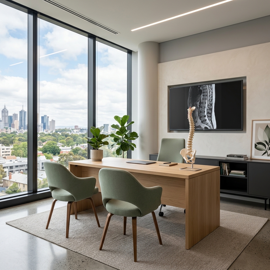
 Clinical anatomical model showing affected spinal structures (no text).
 Causes and risk factors
- Congenital development anomaly where embryonic blood vessel differentiation fails.
- Typically present from birth and matures over time.
- Extremely rare hereditary conditions like Hereditary Haemorrhagic Telangiectasia (HHT) can increase AVM risk.
- Most cases are sporadic and non-hereditary.
How diagnosis is made
- Digital Subtraction Angiography (DSA) is the gold standard for defining AVM anatomy and blood flow.
- MRI and MRA to evaluate the relationship between the AVM and surrounding brain tissue.
- CT and CTA for emergency assessment of acute bleeding.
 Typical diagnostic grey-scale imaging scan (MRI/CT).
 Non-surgical treatment options
- Observation with regular imaging for small, asymptomatic AVMs in deep or highly eloquent brain areas.
- Stereotactic radiosurgery (Gamma Knife) to gradually close the AVM over 2 to 3 years.
- Endovascular embolisation (using medical glue) to reduce blood flow prior to surgery or radiosurgery.
- Anti-seizure medications.
When surgery may be considered
Surgical resection (microsurgical excision) is recommended for AVMs at high risk of rupture or those that have already bled, provided the surgical risk is lower than the natural risk of future bleeding. The goal is complete removal to eliminate rupture risk.
Expected outcomes
Complete surgical resection of an AVM is immediately curative and permanently eliminates the risk of haemorrhage. AVM treatment is highly individualised, combining surgery, embolisation, and radiosurgery for optimal safety.
 Rehabilitation pathways and safe movement restoration.
 Frequently asked questions
How is AVM risk calculated?
The average risk of rupture for an untreated brain AVM is approximately 2% to 4% per year. Each bleeding episode carries a 10% to 15% risk of mortality and a 30% to 50% risk of permanent neurological deficit.