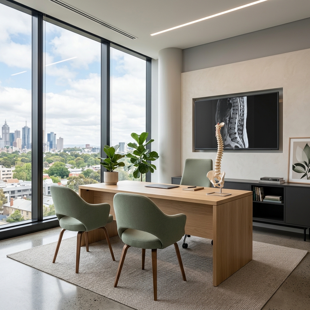
Microdiscectomy
A microdiscectomy is a minimally invasive surgical procedure performed to relieve pressure on a spinal nerve root caused by a herniated disc. Using high-powered operating microscopes and specialized micro-instruments, the surgeon removes only the portion of the ruptured or herniated disc that is compressing the nerve, leaving the rest of the disc intact to maintain structural support and motion.

When this procedure may be recommended
- Severe, radiating leg pain (sciatica) or arm pain that has not responded to conservative treatments (physical therapy, medications, injections).
- Neurological deficits, such as progressive leg weakness or numbness (e.g., foot drop).
- Nerve compression confirmed by MRI matching the clinical symptoms.
- Intolerable pain that severely limits daily function and quality of life.
Who may be a candidate
Ideal candidates are patients with clear evidence of single-level disc herniation causing radicular symptoms (nerve pain) rather than isolated back pain, who have undergone at least 6 weeks of conservative management without sufficient relief, or who present with progressive neurological weakness.
 Advanced medical implant technology (no text).
 Alternatives to surgery
- Structured physical rehabilitation and targeted core-strengthening exercises.
- Analgesic medications, including NSAIDs, neuropathic agents (pregabalin, gabapentin), and oral corticosteroids.
- CT-guided transforaminal epidural corticosteroid injections.
- Alternative minimally invasive procedures, such as endoscopic discectomy.
What to expect
- Anaesthesia & Positioning: The procedure is performed under general anaesthesia with the patient positioned face down on a specialized spinal frame.
- Incision & Access: A small midline incision (typically 1.5 to 2.5 cm) is made in the lower back directly over the herniated disc level.
- Microscopic Visualization: An operating microscope is brought in, providing high-definition illumination and magnification of the spinal canal.
- Nerve Protection: The surgeon gently retracts the compressed nerve root to expose the underlying herniated disc material.
- Disc Removal: The herniated fragment is carefully excised using micro-instruments, relieving pressure on the nerve.
- Closure: The muscles and soft tissues are allowed to fall back into place, and the incision is closed with dissolvable sutures and skin glue.
Technology and imaging
Performed using high-magnification surgical microscopes, high-definition digital visualization, and micro-surgical instrumentation to maximize precision and protect delicate nerve tissues.
 High-precision diagnostic imaging visualization.
  Clinical Zeiss/Leica operating microscope setup.
 Hospital stay
Usually performed as a day procedure, or with a 1-night stay depending on individual patient factors and recovery progress.
Recovery milestones
- Immediate post-op: Mobilisation begins within hours of surgery under physiotherapist guidance.
- Weeks 1-2: Focus on gentle walking, pain management, and wound healing. Avoid bending, lifting, and twisting.
- Weeks 2-6: Gradual increase in walking distance and daily activities. Avoid sitting for longer than 30-45 minutes.
- Weeks 6+: Initiation of structured physical therapy to restore core strength and flexibility. Gradual return to light duties.
Post-operative mobilization and recovery milestones. Risks and complications
- Recurrent disc herniation (5% to 10% risk, occurring in the same disc level).
- Nerve root injury or increased numbness/weakness (less than 1% risk).
- Cerebrospinal fluid (CSF) leak due to dural tear (1% to 2% risk, usually repaired intraoperatively).
- Infection, bleeding, or wound healing complications.
Frequently asked questions
What is the success rate of a microdiscectomy?
Microdiscectomy is highly successful, with about 85% to 95% of patients experiencing significant relief from radiating leg pain (sciatica) almost immediately after the procedure.
When can I return to work after surgery?
For sedentary desk jobs, patients can typically return in 2 to 4 weeks. For physically demanding occupations involving heavy lifting or manual labour, it may take 6 to 12 weeks with light-duty restrictions.
Will I have a large scar?
No. The incision is very small (1.5 to 2.5 cm) and closed with cosmetic techniques, resulting in a minimal, faint scar over time.